

HYPERPLASTIC POLYP OF STOMACH
Dr. Farzana Rahman
Resident, MD Pathology, BSMMU.
Supervisor: Prof. M. Kamal, Chairman, Department of Pathology, BSMMU.
Dr. Farzana Rahman
Resident, MD Pathology, BSMMU.
Supervisor: Prof. M. Kamal, Chairman, Department of Pathology, BSMMU.
CASE REPORT:
Fifty year old female complained of abdominal pain, long lasting dyspepsia and loss of appetite.
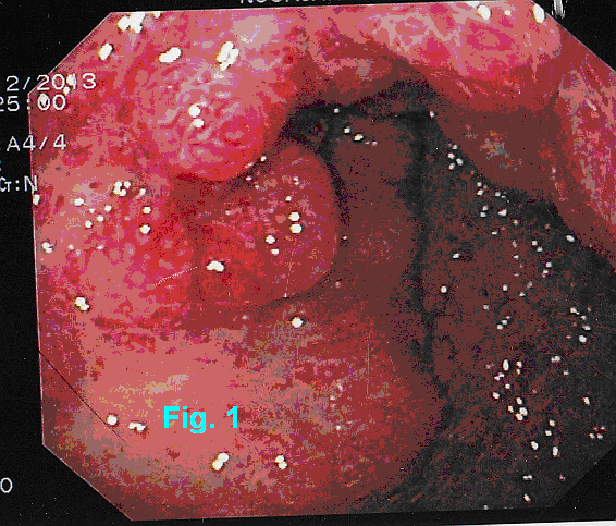
Endoscopic findings of stomach: The mucosa of the bulb was nodular. Nodules were erythematous, more marked at the tip (Fig.1).
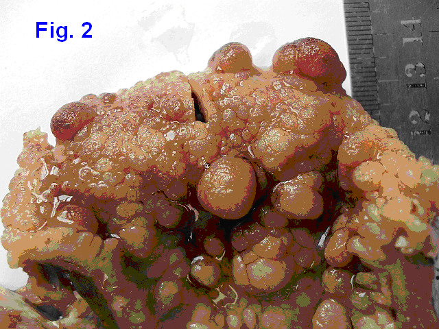
Gross findings : Partial gastrectomy sample, 18 cm along the greater curvature. Multiple sessile polpi in the pyloric end. Largest 1.2 cm (Fig. 2).
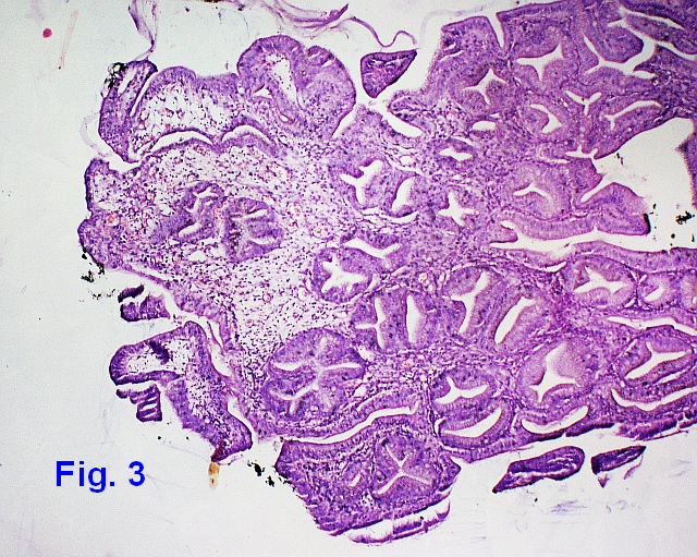
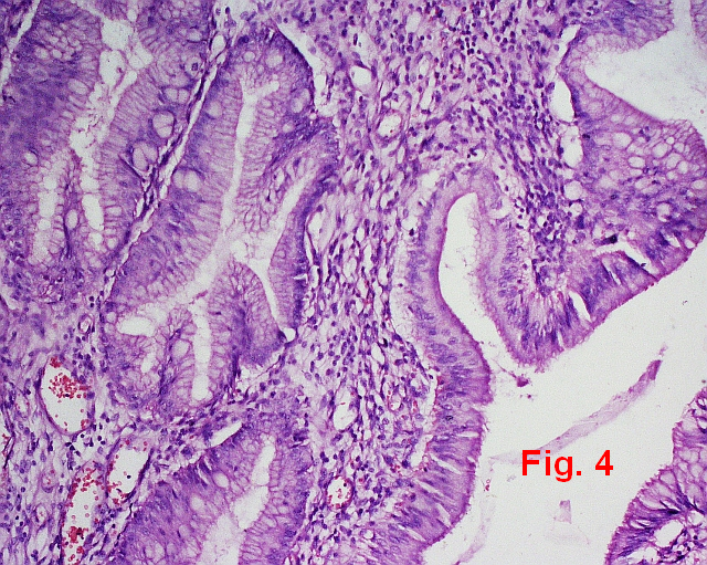
Microscopic features: Irregular, elongated tortuos foveolar glands. The lamina propria is mildly odematous and infiltrated with chronic inflammatory cells. No dysplastic focus was seen. Diagnosis was Hyperplastic polyp of stomach (Fig. 3 and 4).
Fifty year old female complained of abdominal pain, long lasting dyspepsia and loss of appetite.
Endoscopic findings of stomach: The mucosa of the bulb was nodular. Nodules were erythematous, more marked at the tip (Fig.1).
Gross findings : Partial gastrectomy sample, 18 cm along the greater curvature. Multiple sessile polpi in the pyloric end. Largest 1.2 cm (Fig. 2).
Microscopic features: Irregular, elongated tortuos foveolar glands. The lamina propria is mildly odematous and infiltrated with chronic inflammatory cells. No dysplastic focus was seen. Diagnosis was Hyperplastic polyp of stomach (Fig. 3 and 4).
|
|
|
|
Discussion: Also called inflammatory, regenerative polyps, are common gastric polypoid lesions and may approach 75% of all polyps in this site in some population. These are seen on older adults, usually associated gastritis and may be related to H. pylori infection.
Clinical manifestations: Usually asymptomatic and are discovered incidentally on upper endoscopy. They may remain stable, increase in size, or regress following H. pylori eradication
Endoscopic and gross features: Hyperplastic polyps are often multiple and may develop in the antrum, body, fundus or cardia. These are dome-shaped smooth surfaced or may be lobulated. Size range from 0.5 to 1.5 cm. Most are sessile. stalk may be present in a few.
Microscopic features: Composed of elongated, dilated, somewhat distorted crypts lined by foveolar epithelium within an edematous, congested and inflamed lamina propria (Fig. 3). Less common features include goblet cells, intestinal metaplasia, pyloric metaplasia, surface erosion, and ulceration.
Malignancy potentials: Dysplasia may be seen upto 20% cases. Large size (>1cm) and pedunculated polyp carry increased risk of malignancy. Invasive cancer is rare.
Challenges in Gynae and Breast Pathology:
a Festschrift Symposium Honoring Professor K.M. Nazrul Islam
12-13 February 2010
a Festschrift Symposium Honoring Professor K.M. Nazrul Islam
12-13 February 2010
  |
Challenges in Gynae and Breast Pathology:
a Festschrift Symposium Honoring Professor K.M. Nazrul Islam held on 12 and 13 February 2011 12-13 February 2010 Jointly Organized by
Speakers: Prof. W. Dwayne Lawrence MD MSc (Path.) Dr. M. Ruhul Quddus MBBS, M.Phil., MD. |


 |
 |
 |
 |
