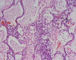
HISTOPATHOLOGY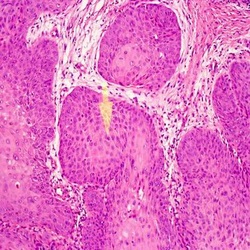
Histopathology service forms the core of patient care and also training of pathology residents. Wide range of specimens are received from BSMMU hospital ans outside. The usual reporting time is 3 days i.e. most of the reports are signed out by 2 p.m. on 3rd day. However, additional time required for special stain(s) and submission of more blocks.
FROZEN SECTION
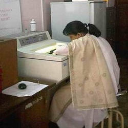
FROZEN SECTION PROCEDURE
Frozen section performed mainly for rapid microscopic analysis. The main steps in this procedure is cryosection –made by cryostat. The report is given within 15- 30 min. It is used for rapid diagnosis e.g. during surgery for intra operative evaluation of the tumour, margins, lymph nodes or other structures such as ganglion cells in Hirschsprung disease. The other uses are in staining tissue where routine processing leads to dissolution or destruction of substances need to examine (e.g. lipid demonstration, Enzyme studies-ATPase , NADPH) and for immunoflurescence studies (Renal and skin biopsy). Frozen section tests done in 2010 at department of Pathology  IMMUNOFLUORESCENCE
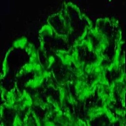
Fluorescence microscopy is done routinely on renal biopsies and requested skin biopsy samples. Tests are done on pooled samples once in a week. One sample is processed for routine stain and a second for direct immunofluorescence staining. For the later fresh samples should be sent in saline soaked gauze.
PHOTOMICROGRAPHY
Photomicrography service is available for academic and research purpose. High quality (8 megapixale) digital photographs are supplied in CD or Pen Drive. Please contact Dr. Nurul Kabir, Assistant Professor, for your requirements.
|
CYTOLOGY
The cytology services offered by the department of Pathology are:
Exfoliative cytology
IMMUNOHISTOCHEMISTRY
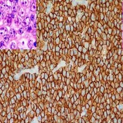
Uses:
1. Categorization of undifferentiated malignant tumours. 2. Determination of site of origin of metastatic tumours. 3. Detection of molecules that have prognotic or therapeutic significance. Guideline for immunohistochemistry test: · Specimen/Paraffin blocks received every weekdays from 8.00 am to 2.30 pm. · Previous histopathology report and clinical information needed. · Report delivered within two weeks (sometime longer period required to issue report in uncommon/difficult cases). Name and price of the Immunohistochemistry markers available at present: 1. ER/PR receptors and Her –2 neu (breast cancer markers) (Tk. 5000) 2. Pancytokeratin, Vimentin, Desmin, S-100 protein, LCA (CD-45) (Markers for undifferentiated tumour) (Tk. 5000) 3. CD3, CD20, CD30. (Marker for lymphoma) (Tk. 5000Tk) 4. CD99 used in Ewing’s sarcoma. (Tk. 2500). 5. CD117 used in GIST. (Tk. 2,500). 6. Chromogranin-A, (Neuroendocrine marker) (Tk. 2,500). 7. GFAP (Used in glioma) (Tk. 2,500) CYTOGENETICS
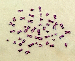
Please contact for karyotyping from pheripharal blood lymphocytes and Barr body analysis. Fresh specimen are required for these tests.

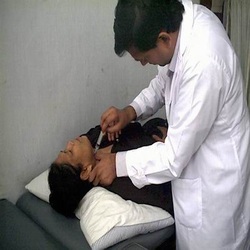
Ultrasound guided FNA is performed regularly. Please make an appointment.
|
|
|
